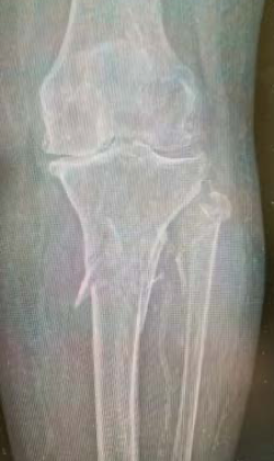
An 83-year-old female living alone at home suffered a fracture to her tibia when she fell out of bed. Based upon the location of the fracture, a suprapatellar approach for the stabilization of the tibia with an IlluminOss implant was made. A small incision and pathway was created above the base of the patella, and a guidewire was introduced. With the guidewire in position, verified by radiographic imaging in both anteroposterior and lateral views, a tissue protector sleeve was inserted over the guidewire, under the patella and positioned on the top of the tibia. A reamer was used to open the medullary canal in the proximal tibia and the canal was prepared for the delivery of an IlluminOss implant.
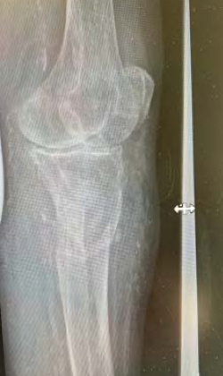
A 22/13mm x160mm implant was chosen as the intramedullary support to the tibia. The delivery sheath was positioned to provide the pathway to the tibial canal and the implant was prepared with monomer. The implant was inserted into the sheath and positioned within the proximal aspect of the tibia under fluoroscopy and then infused with monomer, expanding the implant, and filling the canal. Fluoroscopy was used to ensure that the top of the implant did not protrude or impinge on the articular surface. The implant was cured with visible light, and the catheter was removed with the separation instrumentation.
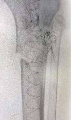
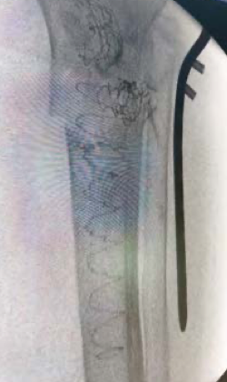
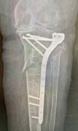
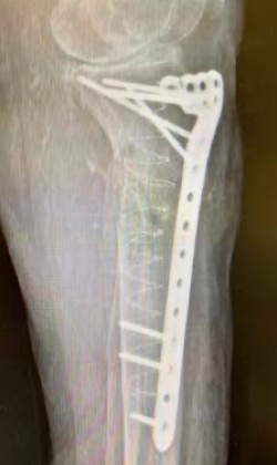
The IlluminOss implant provided strength and stability to the fracture and permitted the planned plate and screws to be securely affixed; as the bone quality was extremely poor, the IlluminOss implant permitted far more screw holding power than the native bone.